


 at screening mammography")
Researchers in Germany say a novel magnetic resonance (MR) breast imaging technique could reduce unnecessary biopsies by providing additional information about suspicious findings on x-ray screening mammography. The technique is non-invasive, does not require a contrast agent and exposes the patient to no ionising radiation. Their work is reported in the journal Radiology.
Conventional x-ray mammography has a high false-positive rate, leading to many unnecessary biopsies. MR imaging could be a useful adjunct to mammography, but the examinations can be time-consuming and commonly require the injection of a contrast agent, which carries its own cost and potential complications.
The researchers evaluated an abbreviated MR breast imaging protocol that requires no contrast agent and uses only two short sequences: the first to show the shape and appearance of the lesion and the second to display its biophysiological properties with diffusion-weighted imaging with background suppression magnetic resonance mammography (DWIBS-MRM). This form of MR imaging works by assessing the diffusion, or movement, of water molecules through tissue. Areas of restricted diffusion may indicate malignancy.
The investigators compared DWIBS-MRM to an abbreviated contrast-enhanced MRI and full diagnostic breast MR protocol in 50 women with suspicious screening mammograms and indication for biopsy. According to the results:
"If the preliminary findings are confirmed, [DWIBS-MRM] could have a high potential to be used as an adjunct in the clarification process of unclear lesions on x-ray mammography in breast cancer screening," says lead author Sebastian Bickelhaupt, MD, a radiologist at the German Cancer Research Center in Heidelberg, Germany. "This might help to reduce the number of invasive biopsies and the related anxiety in women who have suspicious findings at mammography."
With DWIBS-MRM, the MR images can be obtained in less than seven minutes, compared with more than 30 minutes for a full breast MR protocol. The mean reading time using the unenhanced DWIBS-MRM method is less than 30 seconds thanks to an innovative summation technique called maximum intensity projection, or MIP, that allows lesion assessment by reading one summation image instead of multiple single-slice images.
Dr. Bickelhaupt notes that the research is in its early stages and that DWIBS-MRM is not intended as a standalone screening modality but as an adjunct to x-ray mammography and tomosynthesis.
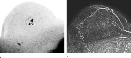
Figure 1. Images show example of a screening-detected lesion in a 67-year-old breast cancer screening patient. (a) Diffusion-weighted imaging with background suppression (b = 1500 sec/mm2) maximum intensity projection (MIP), displayed with black-white inversion, clearly depicts the focal lesion as an area of focal diffusion restriction. (b) First postcontrast subtraction MIP shows an area of subtle enhancement that is partially obscured by generally increased background enhancement.
Source and image credit: RSNA
Conventional x-ray mammography has a high false-positive rate, leading to many unnecessary biopsies. MR imaging could be a useful adjunct to mammography, but the examinations can be time-consuming and commonly require the injection of a contrast agent, which carries its own cost and potential complications.
The researchers evaluated an abbreviated MR breast imaging protocol that requires no contrast agent and uses only two short sequences: the first to show the shape and appearance of the lesion and the second to display its biophysiological properties with diffusion-weighted imaging with background suppression magnetic resonance mammography (DWIBS-MRM). This form of MR imaging works by assessing the diffusion, or movement, of water molecules through tissue. Areas of restricted diffusion may indicate malignancy.
The investigators compared DWIBS-MRM to an abbreviated contrast-enhanced MRI and full diagnostic breast MR protocol in 50 women with suspicious screening mammograms and indication for biopsy. According to the results:
- 24 women had a breast carcinoma
- DWIBS-MRM had a comparable accuracy to that of the other two protocols
- The technique yielded an excellent negative predictive value of 92 percent
"If the preliminary findings are confirmed, [DWIBS-MRM] could have a high potential to be used as an adjunct in the clarification process of unclear lesions on x-ray mammography in breast cancer screening," says lead author Sebastian Bickelhaupt, MD, a radiologist at the German Cancer Research Center in Heidelberg, Germany. "This might help to reduce the number of invasive biopsies and the related anxiety in women who have suspicious findings at mammography."
With DWIBS-MRM, the MR images can be obtained in less than seven minutes, compared with more than 30 minutes for a full breast MR protocol. The mean reading time using the unenhanced DWIBS-MRM method is less than 30 seconds thanks to an innovative summation technique called maximum intensity projection, or MIP, that allows lesion assessment by reading one summation image instead of multiple single-slice images.
Dr. Bickelhaupt notes that the research is in its early stages and that DWIBS-MRM is not intended as a standalone screening modality but as an adjunct to x-ray mammography and tomosynthesis.
* * * * * * * * * *
Top image. Images in 65-year-old breast cancer screening participant with
a suspicious lesion (arrow) at screening mammography. (a) Mediolateral
oblique screening mammogram. (b) Diffusion-weighted imaging with
background suppression (b = 1500 sec/mm2) maximum intensity projection,
displayed with black-white inversion, shows the lesion as an area of
focal diffusion restriction.Figure 1. Images show example of a screening-detected lesion in a 67-year-old breast cancer screening patient. (a) Diffusion-weighted imaging with background suppression (b = 1500 sec/mm2) maximum intensity projection (MIP), displayed with black-white inversion, clearly depicts the focal lesion as an area of focal diffusion restriction. (b) First postcontrast subtraction MIP shows an area of subtle enhancement that is partially obscured by generally increased background enhancement.
Source and image credit: RSNA
Latest Articles
healthmanagement, magnetic resonance, breast cancer screening, mammography, ionising radiation, biopsies
Researchers in Germany say a novel magnetic resonance (MR) breast imaging technique could reduce unnecessary biopsies by providing additional information about suspicious findings on x-ray screening mammography. Their work is reported in the journal R


