Researchers at Florida International University (FIU), Miami, FL, have developed a handheld optical scanner with the potential to offer 3D breast cancer imaging in real time, according to a study published in the journal Biomedical Physics & Engineering Express. The optical device offers several benefits over mammography, with no ionising radiation dose and fewer issues imaging dense tissues.
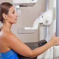
X-ray mammography is the current gold standard for breast cancer detection. This method, however, has a 20 percent false-negative rate (cancer is undetected) and the number increases in younger women with denser breast tissue. Diffuse optical imaging is a safe (nonionising) and relatively inexpensive method for noninvasive imaging of breast cancer.
The new optical scanner uses a near-infrared laser diode source to produce an image of the breast tissues. One advantage presented by the device is that it is more adaptable to breast shape and density. The optical scanner also allows imaging of the chest wall regions, which are harder to image with conventional techniques.
“The women scanned always commented on how comfortable it was to be scanned by our device – many of them said that they didn’t feel anything,” explains Sarah Erickson-Bhatt, PhD, Department of Biomedical Engineering at FIU and an author of the study.
The new scanner builds an image of the tissue by mapping the optical absorption, which is altered by the concentration of haemoglobin – the protein in red blood cells. Regions with higher concentrations of haemoglobin may indicate higher blood floor due to an abnormality such as a tumour.
The research team is currently working on the mathematical tools required to process the images and produce 3D tomographic images, in order to determine tumour size and depth, according to co-author Anu Godavarty, PhD, associate professor, Department of Biomedical Engineering at FIU. “Eventually, we hope that physicians will be able to use this for real-time imaging of breast tissues as part of regular visits by the patients,” says Dr. Godavarty.
The team's ongoing efforts involve extensive clinical work to demonstrate the capability of the device to prescreen for any breast abnormality, followed by seeking FDA approvals prior to clinical use.
Source and image credit: IOP Publishing
References:
Erickson-Bhatt SJ, Godavarty A et al. (2015) Noninvasive surface imaging of breast cancer in humans using a hand-held optical imager. Biomed.
Phys. Eng. Express, 23 October 2015. doi:
http://dx.doi.org/10.1088/2057-1976/1/4/045001
Latest Articles
healthmanagement, breast cancer, mammography, optical imaging, haemoglobin, noninvasive
Researchers at Florida International University (FIU), Miami, FL, have developed a handheld optical scanner with the potential to offer breast cancer imaging in real time, according to a study published in the journal Biomedical Physics & Engineering Expr


