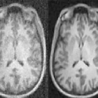
A team of researchers at MIT worked with doctors at Massachusetts General Hospital and other institutions to devise a way to boost the quality of brain scans so that they can be used for large-scale studies of how strokes affect different people and how these people respond to treatment. The paper was presented at the Information Processing in Medical Imaging Conference.
According to Polina Golland, an MIT professor of electrical engineering and computer science, the images used by the researchers are unique because they are acquired in routine practice and are not staged. By using these scans, the researchers are able to study how genetic factors influence survival after stroke as well as how patients respond to different treatments. This approach could also be used to study other disorders such as Alzheimer's disease.
The logic behind using routine scans is simple: these scans are available in vast numbers and provide researchers with a huge set of data. Scientific studies usually require high resolution images but this new approach takes information from the entire set of scans and uses it to recreate anatomical features that are missing from other scans.
"The key idea is to generate an image that is anatomically plausible, and to an algorithm looks like one of those research scans, and is completely consistent with clinical images that were acquired," Golland says. "Once you have that, you can apply every state-of-the-art algorithm that was developed for the beautiful research images and run the same analysis, and get the results as if these were the research images."
By running algorithms that analyse anatomical features and by aligning slices through skull-stripping allows the researchers to retain the information from the original scans but using them as if they were high-resolution images. In other words, the MIT researchers were able to use a technique to enhance low-quality images. They now plan to apply these images to a large set of data to understand spatial patters of damage, disease interaction with cognitive abilities and their ability to recover from the stroke.
"It opens up lots of interesting directions," Golland says. "Images acquired in routine medical practice can give anatomical insight, because we lift them up to that quality that the algorithms can analyse."
Source: Massachusetts Institute of Technology
Image Credit: Massachusetts Institute of Technology
Latest Articles
Stroke, medical imaging, brain scans, skull-stripping
A team of researchers at MIT worked with doctors at Massachusetts General Hospital and other institutions to devise a way to boost the quality of brain scans so that they can be used for large-scale studies of how strokes affect different people and how t


