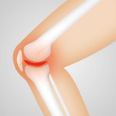
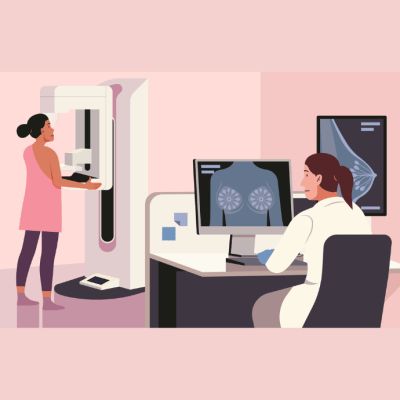
In a study of postmenopausal women with or without mild osteoarthritis, researchers investigated the relation between radiograph-based subchondral bone structure and cartilage composition assessed with delayed gadolinium enhanced magnetic resonance imaging of cartilage (dGEMRIC) and T2 relaxation time. The study findings support the presumption that several tissues are affected in the early osteoarthritis (OA). Further, they indicate that the detailed analysis of radiographs may serve as a complementary imaging tool for OA studies.
OA is considered as a heterogeneous disease which affects all tissues in the joint and has several phenotypes. In the articular cartilage, OA causes progressive degradation and loss of collagens and proteoglycans. OA causes also changes in the density and structure of the subchondral bone. Compositional MRI techniques may be able to capture alterations in the biochemical properties of the tissue already in the early stage of OA. One of the currently available in vivo compositional MRI methods is T2 relaxation time mapping. In the articular cartilage, the integrity and structure of the collagen network and water content affect T2 relaxation time values. Another method is dGEMRIC which has been widely used for the assessment of proteoglycan content of cartilage.
In a previous study, the researchers have shown that bone structure assessed from plain radiographs using Laplacian-based method, local binary patterns (LBP)-based methods, and fractal signature analysis (FSA) is significantly related with the actual 3-D microstructure of tibial bone. They have also reported that subchondral and trabecular bone structures evaluated using LBP-based and Laplacian-based methods differ between subjects with different Kellgren–Lawrence (KL) grades.
In the current study, the researchers sought to assess the sensitivity of the radiograph-based structural analysis of bone to early OA by comparing the methods to compositional MRI. The study included 93 postmenopausal women (Kellgren–Lawrence grade 0: n = 13, 1: n = 26, 2: n = 54). Radiograph-based bone structure was assessed using entropy of the Laplacian-based image and local binary patterns, homogeneity indices of the local angles, and horizontal and vertical fractal dimensions. Mean dGEMRIC index and T2 relaxation time of tibial cartilage were calculated to estimate cartilage composition.
The results showed several statistically significant correlations between different radiograph-based bone structure-related parameters and cartilage composition assessed with dGEMRIC and T2 relaxation time. "The direction (positive/negative) of the correlations indicates that when tibial cartilage is degenerated, the underlying tibial bone structure is also deteriorated," the researchers explain.
However, the relation between subchondral bone structure and composition of cartilage was not very strong and perfectly linear, which was expected, according to the research team. One possible explanation for the relatively low correlations is that OA is a heterogeneous disease and likely has different origins.
"The selection of subjects with different OA phenotypes is challenging and it is probable that many phenotypes were mixed in our sample. Furthermore, it may be that not all tissues are affected in the early stage of the disease which may affect the correlation levels. Moreover, we investigated different tissues, i.e., cartilage using MRI and bone using radiographs, but the interplay between subchondral bone and articular cartilage is not clear yet. Estimation of the clinical significance of the results is challenging," the authors write.
Although statistically significant correlations between MRI-based cartilage composition and radiograph-based subchondral bone structure were observed, the authors say more detailed studies with carefully selected subjects (e.g., subjects that have or are at risk of developing bone changes) and imaging modalities are needed in order to assess clinical relevance of the results and to further understand how and which factors affect the changes in the cartilage and subchondral bone.
Source: Osteoarthritis and Cartilage
Image Credit: Pixabay
Latest Articles
osteoarthritis, radiography, postmenopausal women
In a study of postmenopausal women with or without mild osteoarthritis, researchers investigated the relation between radiograph-based subchondral bone structure and cartilage composition assessed with delayed gadolinium enhanced magnetic resonance imagin

