Researchers at Lund University in Sweden
and the Massachusetts Institute of Technology in the U.S. have developed a
method called iso-acoustic focusing to analyse and separate cells from the
blood. Ultimately, this method, can become significant to measure the
efficiency of cancer treatments for individuals, according to their research
published in Nature
Communications.
In brief, the new method involves exposing cells to ultrasound when they flow through a so-called micro-channel inside a chip. The individual cells are separated in the acoustic field and by studying the cells’ lateral movement at the end of the channel it is possible to identify the acoustic properties of the cells. Conversely, if you know the cells’ acoustic characteristics, you can detect which type of cell that passes through.
“The vision is that our innovation will eventually be used in healthcare facilities, for example, to count and distinguish different types of cells in patients’ blood”, said Per Augustsson, researcher at the Department of Biomedical Engineering at Lund who developed the method together with researchers at the Technical University of Denmark during his time as postdoc in the lab of Professor Joel Voldman at M.I.T., with funding from the Swedish Research Council.
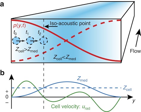
(a) Cells (circles) flowing in a microchannel are deflected sideways towards the node of an acoustic resonant pressure field p (red curves) in a medium of position-dependent acoustic impedance Zmed (color plot from low (white) to high (blue)). (b) Conceptual plot showing that when the acoustic impedance Zcell (dashed blue line) of a given cell matches Zmed (full blue line) at the IAP, its transverse velocity urad (green line) becomes zero so that its position along y reflects its individual effective acoustic impedance.
The researchers have applied the method to measure the acoustic properties of white blood cells and discovered that there are differences between different subgroups. Furthermore, the researchers have observed that cancer cells that are cultured in a laboratory have significantly different acoustic properties in comparison with blood cells from healthy donors.
“It may seem odd that we are interested in the acoustic properties of blood cells and cancer cells. But we have been searching for new methods to separate cells in order to study them in more detail”, said Augustsson.
The blood contains extremely rare cells and it can be of interest to gain access to them. One example is the so-called circulating tumour cells found in the blood of patients with cancer, which play a major part in the spread of cancer inside the body.
The ability to measure how the number of tumour cells varies from one occasion to another can help determine whether medication in the context of a treatment will have the desired effect. But this is technically very challenging and research groups worldwide are currently developing methods to achieve this.
One strategy is to employ acoustic fields. In previous studies in acoustic separation of cells it has only been possible to separate the cells based on their size. However, measuring the size of cells is in itself not enough to determine the cell type in question.
“Since we are looking for individual cells in a blood sample which contains billions of cells, the smallest overlap in size between the cancer cell and other blood cells will lead to thousands of blood cells ‘contaminating’ the cancer cells extracted through the separation. This is why we have now developed iso-acoustic focusing”, added Augustsson.
The new method provides a way to count and measure the acoustic-mechanic properties of the cells, and the hope is that in the future this will create a better understanding of, for example, how cancer spreads in the body. With the help of this method, researchers hope to shed light on issues such as: What causes metastasis, and which mechanisms control how tumour cells spread in the body? Are there differences in physical characteristics between tumour cells and circulating tumour cells?
“We are currently also working on two follow-up projects to describe the physics behind iso-acoustic focusing in greater detail. This is a very exciting project, run by myself together with physics professor Henrik Bruus and doctoral student Jonas Tobias Karlsen, both at the Technical University of Denmark. We expect that the work will lead to meaningful results within our field of research – acoustofluidics”, concluded Augustsson.
Reference:
Full bibliographic information Augustsson P. et al (2016). Iso-acoustic focusing of cells for size-insensitive acousto-mechanical phenotyping. Published in Nature Communications; doi:10.1038/ncomms11556