Researchers at The University of Nottingham have developed a new test for the diagnosis of Parkinson’s Disease using standard scanning equipment common in clinical offices. 3T MRI is employed to scan sub-regions of the brain in search of a group of cells shaped like the tail of a swallow. The image is present in the scans of brains not affected by Parkinson’s Disease, but absent in those of patients diagnosed with the degenerative condition.
The research findings, published without restricted access in PLOS one, document the work of neuroradiology experts Dr. Stefan Schwarz and Professor Dorothee Auer of the School of Medicine at The University of Nottingham. Dr. Nin Bajaj of the Nottingham University Hospitals NHS Trust contributed expertise in movement disorder diseases. The work was carried out at the Queen's Medical Centre and builds upon a collaboration with Professor Penny Gowland of the Sir Peter Mansfield Magnetic Resonance Centre at The University of Nottingham.
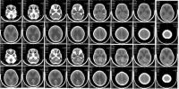
A ‘Tell-Tail’ MRI Image
The study describes a distinctive image in the shape of a swallow’s tail, which is attributable to healthy sub-regions of the brain. In Parkinson’s Disease, there is a loss of substantia nigra dopaminergic neurones, which are most prominent in parts of the brain known as nigrosomes. High resolution MRI permits direct visualisation of such tissue in the brain scans of healthy people. Depletion of the neurones in Parkinson’s Disease leads to the absence of such imagery during magnetic resonance imaging.
Parkinson's Disease is characterised by a progressive destruction of brain cells responsible for controlling movement. Symptoms include tremor and rigidity. Approximately 127,000 people suffer from the condition in the UK. There is currently no cure for the disease, although drugs are available to control some symptoms.
Diagnostic Challenges
Compared to existing diagnostic techniques, the new test is considerably less costly and remarkably accurate. Parkinson’s Disease is notoriously difficult to diagnose in vivo, particularly in its early stages, and clinical certainty often depends upon the expensive and potentially dangerous involvement of nuclear medicine. Non-licensed diagnostic methods are unreliable and vary in their accuracy and ease of replication, ruling them out for clinical practice.
According to Dr. Schwarz, "This is a breakthrough finding as currently Parkinson's Disease is mostly diagnosed by identifying symptoms like stiffness and tremor. Imaging tests to confirm the diagnosis are limited to expensive nuclear medical techniques which are not widely available and associated with potentially harmful ionizing radiation.”
New Hope with Existing Equipment
High resolution MRI with ultra high field 7T imaging allowed the Nottingham researchers to identify the peculiar pathology of Parkinson’s Disease. The condition involves structural changes to the mid brain in a small area called the substantia nigra. Following a review of 114 high resolution scans, the team accurately diagnosed 94% of the cases. Furthermore, their findings indicate that widely accessible 3T MRI scanners are capable of detecting the brain changes. This means that many hospitals across the world already possess the only necessary tool for carrying out the new test.
"Using Magnetic Resonance Imaging (no ionizing radiation involved and much cheaper than nuclear medical techniques) we identified a specific imaging feature which has great similarity to a tail of a swallow and therefore decided to call it the 'swallow tail sign'. This sign is absent in Parkinson's disease," said Dr. Schwarz.
'The 'Swallow Tail' Appearance of the Healthy Nigrosome - A New Accurate Test of Parkinson's Disease: A Case-Control and Retrospective Cross-Sectional MRI Study at 3T' is freely available to the public in the academic journal PLOS one.
References:
Schwarz ST, Afzal M, Morgan PS, Bajaj N, Gowland PA, et al. (2014) The ‘Swallow Tail’ Appearance of the Healthy Nigrosome – A New Accurate Test of Parkinson's Disease: A Case-Control and Retrospective Cross-Sectional MRI Study at 3T. PLoS ONE 9(4): e93814. doi:10.1371/journal.pone.0093814
Latest Articles
Imaging, MRI, brain atrophy, Parkinsons' Disease, diagnostic imaging, high resolution MRI, brain cells
Researchers at The University of Nottingham have developed a new test for the diagnosis of Parkinson’s Disease using standard scanning equipment common i...