


1. Background:
Extensive clinical diagnosis experience has been obtained within the field of myocardium disease, coronary disease, valve disease, cardiac tumor and congenital disease by echo practitioners, making echocardiography to emerge as a rather common practice within the last couple of decades. Technologies such as B-mode, Mmode, Color Doppler etc. continue to contribute in the improvements within echocardiography. However, B-mode, as a robust and direct anatomical visualization technology, remains to be the most important one in echocardiography.
Ultrasound waves used for echo imaging consist of main lobe and side lobe. Although side lobe is much weaker than main lobe, when side lobe propagates through hyperechogenic region (pericardium or free air bubbles), it generates useless clutter which is much stronger than real echo, leading to creation of noise in the chambers.
During a regular echo scan, ultrasound probe is usually placed at the parasternal area or the fourth intercostals. For patients with wider intercostals space, ultrasound waves are easier to penetrate into the tissue. However, for patients with smaller intercostals space, part of ultrasound waves transmitted or echo signals are blocked by ribs due to its high density and strong attenuation, forming a blind area underneath the ribs. Hence, cardiac tissue located within the blind area tends to be of low S/N ratio, resulting in less information being obtained from the region.
Due to different body types and attenuation rate, signals transmitted in tissue vary from person to person. What’s more note worthy for echocardiography, is that the heart and chest movement can make a case more complicated than any other organ. For instance, an eco scan conducted on one patient, at same position, orientated at same view, can result in different images being obtained from time to time. The distribution of signal intensity differs depending on the body type, time and position. To get better image quality, it is necessary to optimize imaging parameters for different cases.
2. Echo Boost Introduction:
Utilizing a unique intelligent data receptive method for echo data analysis for specific region, Echo Boost is a fully self-adaptive signal processing technology, designed to optimize whole field uniformity of myocardium and chamber.
Echo Boost uses a receptive field technology to get echo data. Receptive field originates from neurology, in which neurons are activated as a result of stimulation in a specific region, however, for ultrasound; it applies to auto adaptive reception for echo received. For instance, in Graphic 1, waves are relatively high intensity echo “Echo1”, “Echo2”, “Echo3”, and other dash lines indicate low echo region such as chamber. Horizontal and vertical lines specify the location of receptive field and intensity distribution of the receptive field respectively.
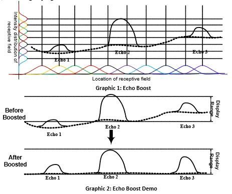
Location of Receptive Field divides a region into dozens of smaller images, and analyzes each region individually. During processing, the center of region is very active, and the outer area of the region suppresses the echo reception.
Intensity Receptive Field detects intensity distribution, and estimates current echo signals, obtaining any signal characteristic in current receptive field. The signal is detected not only from spatial distribution, but also from intensity, covering whole range of echo signals, with ability to suppress noise and boost useful weak signals.
When receptive field signal characteristic acquisition is finished, ehco signals are processed as illustrated below: for weak echo “Echo1”, the signal is boosted to a user visible range; for stronger signal “Echo2”, the signal is compressed to avoid to be too bright to increase detail info; for “Echo3”, part of gain and contrast is amended, keeping homogeneity with improved “Echo1” and “Echo2”.
3. Benefits:
a) Significantly improves B-mode whole field uniformity of cardiac image
b) Significantly improves contrast resolution of cardiac tissue and chambers, reducing chamber noise.
4. Case Study:
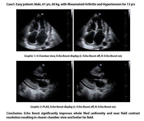
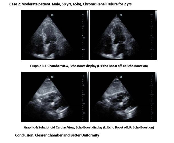
A series of echo examinations were conducted on 3 different patients to compare the result and test the validity of the Echo Boost technology. It can be concluded that Echo Boost can effectively improve the uniformity and contrast of an image and at the same time reduce the noise especially in chamber for all body types.
Latest Articles
Ultrasound, Technology, echocardiography, Mindray
1. Background: Extensive clinical diagnosis experience has been obtained within the field of myocardium disease, coronary disease, valve disease, cardiac t...


