A large consortium of European researchers headed by the University of Twente (UT) will receive grants of over five million euros to develop a new imaging device for the diagnosis of breast cancer. The device prototype will be ready for testing and production in four years.
The new device is expected to offer improved photoacoustic and ultrasound imaging and will be used to combine images that are generated by both techniques. It is expected that this new imager will help improve and accelerate breast cancer diagnosis and can also be used effectively in younger women.
Currently, the techniques used for breast cancer diagnosis such as x-ray mammography, ultrasound and MRIs have shortcomings. The biggest disadvantage with these techniques is the fact that they cannot distinguish between a tumour from healthy tissue or a benign abnormality. This often results in missed tumours and the need to carry out biopsies.
Researchers will be developing a completely new device that will shorten the time to accurate diagnosis and will be especially useful among young women where x-ray mammogrphy generally does not perform well. No radiation or contract media will be used with this device and there will no pain caused to the women who are examined with it.
The European Union has granted 4.35 million Euros for the development of this device. The remaining balance will be provided by the Swiss government. During the development of the device, dubbed PAMMOTH, a dream imager, input and advice about design and testing will be also be taken from doctor and patient associations.
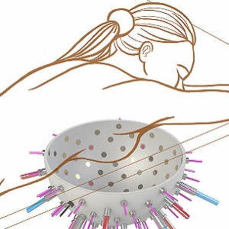
Project leader Srirang Manohar explains that it will combine photacoustic and ultrasound imaging techniques. Photoacoustic mammography (PAMmography) illuminates the breast with a laser light which is then converted into ultrasound in regions with higher concentration of blood such as the areas around a malignant tumour. The ultrasound produced thus travels from the tumour to the skin where it can be easily detected. In addition, the researchers will develop technology that will generate a three-dimensional image.
“The images from the two systems will be combined together.
This will result in simultaneous three-dimensional information about
disease specific optical contrast, as well as about the ultrasound
properties which provide anatomic information within the breast. Further
we wish to do this combined imaging in real time," said Manohar.
Source: University of Twente
Image Credit: Srirang Manohar


