HealthManagement, Volume 18 - Issue 2, 2018
 PRINT OPTIMISED
Radiologists collaborate with sports physicians for optimal performance
How diagnostic imaging can be used in sports medicine and how different techniques can be used to assess muscle injuries.
Diagnostic imaging in sports medicine
As
muscle injuries represent more than thirty percent of sports injuries,
with great implications for professional teams, musculoskeletal
radiologists need to share the new knowledge of sports medicine in terms
of diagnosis, prognosis and return to play in muscle injuries.
New
injury classifications have been published, in order to connect the
imaging findings and clinical examination and thus improve the grade of
severity of muscle injuries and the possibility of re-injuries.
It is
very important to get the best diagnosis after muscle injuries in order
to determine the severity of the injury (in other words the time for
return to play/ sport/competition) and the risk of re-injury.
For this reason, cross-collaboration is of key importance between:
- Sports physicians with vast experience in muscle injuries and return to play together with expertise in ultrasound
- Musculoskeletal radiologists with a background in muscle injuries and expertise in new imaging sequences and 3 Tesla magnetic resonance imaging (MRI)
Ultrasonography and magnetic resonance imaging are
currently the most frequently applied techniques in sports medicine.
Imaging is crucial to confirm and assess the extent of sports-related
muscle injuries and may help to guide management, which directly affects
the prognosis. Prognosis based on the available clinical and imaging
information is vital.
Classification of muscles injuries
Although
different classification systems have been published, because they are
used in daily practice, it may be challenging to classify injuries due
to varying muscle architecture, lesion extension, activities when the
lesion occurred and even the mechanism.
Recently three new classifications have been proposed:
1. Munich Consensus Classification (Mueller- Wohlfahrt et al. 2013)
2. British Classification System (pollock et al. 2014)
3. FCB Barcelona and Aspetar Classification (Valle et al. 2017)
These classifications focus on different factors that are key for a best diagnosis and prognosis:
1. Mechanism of injury
2. Location of the injury
3. If myofascial, myotendinous or intramuscular tendon affected
4. If there is retraction of fibres or not
5. Extension of oedema (& cross-sectional area)
6. If it is a re-injury
These
classification systems are based on the current available research and
experience of clinical experts from different institutions with
experience in assessing a high volume of muscle injuries.
However, we
need a consensus to improve communication between all sports
physicians and radiologists in athlete-related professional
relationships regarding muscle injuries.
How MRI findings contribute to diagnosis and prognosis
3 Tesla MRI techniques have contributed to getting better images of the muscle architecture and muscle injuries, especially the level and quantity of myoconnective tissue. this means that the extracellular matrix involved might be variable, based on the principle that the more connective tissue is damaged, the greater the functional impairment—and worse prognosis.Currently we are able to read how muscle injuries affect the myoconnective tissue, myofascia, i.e. more peripheral injury, myotendinous, i.e. typical muscle injury with feather imaging as well as intramuscular tendon injury. We have clinical evidence that when an injury has affected the intramuscular tendon, such as the common biceps/semitendinosus tendon, the prognosis was far worse and the incidence rate of an injury was higher.
When the doctor identifies a gap or retraction of muscle
fibres, it shows there is a criterion for bad prognosis—a “marker” with
less scientific evidence. In practical terms this means that the
severity of a muscle injury in the same location can depend on the
different histoarchitecture alteration, i.e. the difference between an
injury that can last weeks or needs to be prescribed surgical
treatment.
Predicting return to play and re-injury risk At
least for now there is no strong evidence that a 1.5 Tesla MRI finding
is useful for predicting the time to return to sports. There is evidence
in the sports medicine world that normalisation of increased signal
intensities on MR images is not required for a successful return to
play, suggesting that functional recovery precedes structural recovery
on imaging.
demonstrate that the first criterion for return to play is a positive diagnosis and everyday imaging techniques would indeed help us. Secondly, in the rehabilitation process if something occurs with an individual, we can monitor the response using both ultrasound (US) and MRI, together with monitoring of pain, flexibility, fatigue and strength.
In the future, there will no doubt be new evidence and we will be able to to focus on areas such as:
• How fibrosis in previous injury has affected the recover y process, seen on t1-weighted Mri images
• How elastography could help as a marker of flexibility
• How we can see changes in the architecture remodelling using diffusion tensor imaging
• How the healing process by t2-weighted mapping is changing in terms of recruitment of muscles
Ultrasound, at least in our experience, is good for monitoring the healing process and controlling haematoma, fibrosis, architecture repair etc. It is cheaper and more useful but it’s certainly not the best return-to-play marker decision.
Another frequent question in our field is whether Mr imaging findings allow us to predict re-injury? the answer is not for now—there is no great evidence to support the use of Mr imaging findings to assess extent of injury or histoarchitecture-affected fibrosis for identifying athletes at increased risk of re-injury. However, our impression is that it will take place, but more research is needed.
Many sports medicine physicians have learned to appreciate high-quality imaging to help guide athletes in recovery processes, although the clinical evaluation itself and other tools such as GPS values, must
guide the final Return to Play decision, at least at the moment.
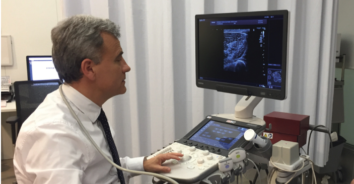
Ultrasound imaging of muscle injuries these
days, at least in Spain, using ultrasound (US) is similar to a
cardiologist using a stethoscope. We always diagnose muscle injuries
with the support of ultrasound. this requires vast experience, but it is
really useful and we encourage specialists from other countries to also
use this tool.
In our opinion, we agree that using
ultrasound is less sensitive than MR imaging, but on the other hand with
US you can observe and monitor very easily. Soon the evolution of the
healing process, identifying if haematoma persists, fibrosis appears
and if the fibrillary architecture is remodelling in the right way, will
become clear. this is naturally really useful for daily practice.
Another
advantage of using ultrasound for the evaluation of healing in muscle
injuries is the ability to perform a dynamic assessment before and after
muscle contraction, which may or may not depict persistence of fibre
disruption, after clinical management and rehabilitation. This is very
important in the evaluation of moderate to severe muscle injuries,
which are the reasons why Return to Play may be delayed.
Future techniques
Advanced MR imaging techniques available for muscle assessment are not applied routinely in clinical practice. but it’s true that professional medical staff are promoting and asking radiologists for new techniques for better diagnosis and prognosis.For example:
- T2-weighted mapping may be useful from a sports medicine perspective. T2 values increase in stressed muscles and can help us know activation or changes in muscle recruitment after muscle injuries
- Diffusion tensor imaging allows diffusion quantification of anisotropic tissues and allows us to see muscle fibre direction tracking, to detect minor muscle injury and to differentiate injured muscles from normal.
- Skeletal muscle MR elastography can be used for studying the physiologic response of normal or damaged muscles. In fact, it has been found that there is a difference in the stiffness of muscles after injury.
Key points
- Muscle injuries represent more than thirty percent of sports injuries
- Optimal diagnosis of muscle injury deter- mines the severity of the injury and time to return to play and risk of re-injury
- Cross-collaboration between sports physicians and musculoskletal radiologists is of key importance
- US and MRI are the most used techniques in sports medicine
- In future, T2-weighted mapping, DTI, MR elastography and PET are expected to play a greater role as more research on these is done
References:
Mueller-Wohlfahrt HW et al. (2013) Terminology and classification of muscle injuries in sport: The Munich consensus statement. Br J Sports Med 47(6): 342-50.
Pollock N et al. (2014) British athletics muscle injury classification: a new grading system. Br J Sports Med 48(18): 1347-51.
Valle X et al. (2017) Muscle injuries in sports: a new evidence-informed and expert consensus-based classification with clinical application. Sports Med 47(7): 1241-53.
Yamada AF et al. (2017) Diagnostic imaging of muscle injuries in sports medicine: new concepts and radiological approach. Curr Radiol Rep 5: 27.