New research from Thomas Jefferson University suggests that contrast-enhanced subharmonic imaging (SHI) could be useful in detecting prostate cancers that are not identified by MRI. The findings will be presented at the ARRS 2018 Annual Meeting, set for 22-27 April in Washington, DC.
SHI is a new technique for imaging of microbubble ultrasound contrast agents with improved suppression of background tissues. The latest findings indicate that SHI provides improved conspicuity of microbubble contrast enhancement and may improve the performance of contrast-enhanced ultrasound for detection of prostate cancer.
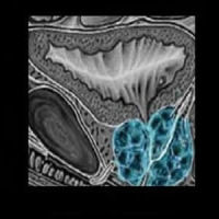
The research is the first in vivo application of contrast-enhanced SHI for the detection of prostate cancer. Among the study group of 28 patients, contrast enhancement was clearly observed with colour and power Doppler imaging, as well as with both harmonic imaging (HI) and SHI techniques in all subjects. SHI provided improved contrast signal and tissue suppression relative to conventional HI. Areas of increased vascularity were best delineated with maximum-intensity projection SHI, which allowed visualisation of microvascular architecture.
Notably, contrast-enhanced SHI demonstrated contrast enhancement in the prostate of every study patient, with focal areas of contrast enhancement corresponding to sites of cancer in 18 percent of targeted biopsies, including five patients whose cancer was not identified by MRI, according to researchers.
Venkata Masarapu of Jefferson Medical College of Thomas Jefferson University will present the results of the study at the ARRS 2018, which has lined up educational activities representing the entire spectrum of radiology. Leading radiologists from around the world are expected to attend the annual meeting to be held at the Marriott Wardman Park Hotel in Washington, DC.
Source: American Roentgen Ray Society (ARRS)
Image Credit: Wikimedia Commons
Latest Articles
prostate cancer, Contrast-enhanced ultrasound, contrast-enhanced subharmonic imaging, MRI.
New research from Thomas Jefferson University suggests that contrast-enhanced subharmonic imaging (SHI) could be useful in detecting prostate cancers that are not identified by MRI. The findings will be presented at the ARRS 2018 Annual Meeting, set for