


According to a new study, magnetic resonance imaging (MRI) may identify additional breast cancers that are sometimes potentially more aggressive than those found on mammography. Researchers say that, in some cases, MRI findings of additional cancers not seen on mammography may necessitate a change in treatment. The results are published in the journal Radiology.
As the most sensitive technique for detecting breast cancer, MRI is effective in the detection of what are known as multicentric cancers — ie, breast cancers involving two or more distinct primary tumours, usually in different quadrants of the breast. However, the clinical significance of multicentric cancers found with MRI has been the subject of ongoing debate.
“Patients with clinically insignificant cancers undergoing potential overtreatment versus patients who may be undertreated is at the heart of the controversy surrounding breast MRI,” says lead author Chiara Iacconi, MD, from the Breast Unit at USL1 Massa-Carrara in Carrara, Italy.
For this study, the researchers reviewed records from 2,021 patients with newly diagnosed breast cancer who underwent biopsy after preoperative MRI. Of these patients, 285 (14 percent) had additional cancer detected on MRI that was hidden from view on mammography. In 73 (25.6 percent) of those 285 patients, MRI identified at least one additional cancer in a different quadrant of the breast than the index cancer — ie, the cancer detected by mammography and/or breast palpation.
Notably, these multicentric cancers were larger than the known index cancer in 17 (23.3 percent) of the 73 patients. In addition, the MRI-detected multicentric cancers were greater than 1 cm in size in 25 percent of the 73 patients.
The additional MRI-detected multicentric cancers were found mostly in patients with heterogeneously dense or extremely dense breasts, the researchers point out. However, MRI also detected additional disease in 19 percent of patients with fatty or scattered fibroglandular tissue.
“The results show that multicentric cancer detected on breast MRI after mammography appears to represent a larger tumour burden in approximately a quarter of patients and can result in potential changes to cancer grade and treatment,” says Dr. Iacconi.
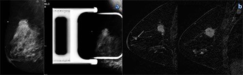
Figure 1. Mammography and MR imaging in a 46-year-old woman with a
palpable mass in the right breast. A: Mediolateral oblique (left) and
spot compression (right) mammograms in the right breast show a mass in
the upper-outer quadrant. B: Sagittal dynamic breast MR images in the
right breast show an en-hancing mass with speculated, or spiky, margins
in the right upper outer quadrant, an additional focus near the nipple
(DCIS), and nonmass enhancement in the inferior quadrants (DCIS). The
additional lesions were not visible at US and were biopsied with MR
imaging guidance.
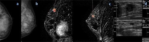
Figure 2. Mammography and MR imaging in a
48-year-old woman with high risk for breast cancer. A: Screening
mammogram shows a craniocaudal view of the left breast. B: Mediolateral
oblique screening mammogram (left) shows the left breast, and dynamic
sagittal MR image (right) of the left breast shows a 1.3-cm index lesion
(grade I CDI and grade II DCIS). C: dynamic sagittal MR image shows an
additional 1-cm lesion (grade II CLI and grade III DCIS). D: US image
shows both lesions and was used to guide biopsy.
Source: Radiological Society of North America (RSNA)
Image credit: RSNA; Flickr.com
As the most sensitive technique for detecting breast cancer, MRI is effective in the detection of what are known as multicentric cancers — ie, breast cancers involving two or more distinct primary tumours, usually in different quadrants of the breast. However, the clinical significance of multicentric cancers found with MRI has been the subject of ongoing debate.
“Patients with clinically insignificant cancers undergoing potential overtreatment versus patients who may be undertreated is at the heart of the controversy surrounding breast MRI,” says lead author Chiara Iacconi, MD, from the Breast Unit at USL1 Massa-Carrara in Carrara, Italy.
For this study, the researchers reviewed records from 2,021 patients with newly diagnosed breast cancer who underwent biopsy after preoperative MRI. Of these patients, 285 (14 percent) had additional cancer detected on MRI that was hidden from view on mammography. In 73 (25.6 percent) of those 285 patients, MRI identified at least one additional cancer in a different quadrant of the breast than the index cancer — ie, the cancer detected by mammography and/or breast palpation.
Notably, these multicentric cancers were larger than the known index cancer in 17 (23.3 percent) of the 73 patients. In addition, the MRI-detected multicentric cancers were greater than 1 cm in size in 25 percent of the 73 patients.
The additional MRI-detected multicentric cancers were found mostly in patients with heterogeneously dense or extremely dense breasts, the researchers point out. However, MRI also detected additional disease in 19 percent of patients with fatty or scattered fibroglandular tissue.
“The results show that multicentric cancer detected on breast MRI after mammography appears to represent a larger tumour burden in approximately a quarter of patients and can result in potential changes to cancer grade and treatment,” says Dr. Iacconi.
Source: Radiological Society of North America (RSNA)
Image credit: RSNA; Flickr.com
Latest Articles
healthmanagement, breast cancer, breast MRI, biopsy, mammography, primary tumour
According to a new study, magnetic resonance imaging (MRI) may identify additional breast cancers that are sometimes potentially more aggressive than those found on mammography.
, scattered fibroglandular (top right), heterogeneously dense (bottom left) and extremely dense (bottom right).")


