


A brain imaging study, due to be published in PLOS One, has shown that, compared to healthy control subjects, patients with chronic fatigue syndrome (CFS) (also known as myalgic encephalomyelitis or ME) may have reduced responses in a region of the brain connected with fatigue. This suggests that chronic fatigue syndrome is associated with changes in the brain involving brain circuits that regulate motor activity and motivation.
As measured by fMRI (functional magnetic resonance imaging), patients with CFS had less activation of the basal ganglia, and this was linked with the severity of fatigue symptoms.
CFS is a debilitating and complex disorder. Patients experience intense fatigue that is not improved by bed rest, and that may be worsened by exercise or mental stress.
Lead author Andrew Miller said that they chose the basal ganglia, because they are primary targets of inflammation in the brain, and previous studies suggest that increased inflammation may be a contributing factor to fatigue and even the cause in CFS patients.
The study was a collaboration among researchers at Emory University School of Medicine, the U.S. Centers for Disease Control and Prevention's Chronic Viral Diseases Branch, and the University of Modena and Reggio Emilia in Italy.
The basal ganglia, situated deep in the brain, are thought to be responsible for control of movements and responses to rewards as well as cognitive functions. Neurological disorders that involve dysfunction of the basal ganglia include Parkinson's disease and Huntington's disease.
Miller noted that previous studies have suggested that responses to viruses may underlie some cases of CFS and added that the data in this study support the idea that the body's immune response to viruses could be associated with fatigue by affecting the brain through inflammation.
The study compared 18 patients diagnosed with chronic fatigue syndrome with 41 healthy volunteers. The study excluded anyone with depression or on antidepressants, but not those with anxiety disorders.
For the brain imaging portion of the study, participants were told that if they correctly guessed whether a preselected card was red or black they would win a dollar. After their guess, the colour of the card was revealed, and blood flow to the basal ganglia was measured.
The key measurement was the difference in activity between a win or loss. Participants' scores on a survey measuring their levels of fatigue were tied to the difference in basal ganglia activity between winning and losing. Those experiencing most fatigue had the smallest changes, especially in the right caudate and the right globus pallidus of the basal ganglia.
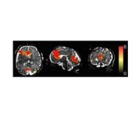
Image caption:
Basal Ganglia Activation in the Gambling Task.
Left to right - Axial, coronal and transverse sections of the brain. The top row displays the activation for the Win-Lose contrast, for the pooled sample of chronic fatigue syndrome (CFS)+Control subjects, as a statistical parametric map thresholded at a p<0.05 corrected threshold and masked with the atlas-based anatomical regions of interest portrayed in the bottom row (putamen: purple; caudate: orange; globus pallidus: turquoise).
doi:10.1371/journal.pone.0098156.g001
Source: EurekAlert
References:
Miller AH, Jones JF, Drake DF et al. (2014) Decreased basal ganglia activation in subjects with chronic fatigue syndrome: association with symptoms of fatigue. PLoS ONE 9(5): e98156. doi:10.1371/journal.pone.0098156
Latest Articles
functional MRI, chronic fatigue syndrome
A brain imaging study, due to be published in PLOS One, has shown that, compared to healthy control subjects, patients with chronic fatigue syndrome (CFS)...

 patients and, B, exclusively in PD + visual hallucinations (VH) patients. Regional functional connectivity analysis revealed lower functional connectivity in PD + VH a")


