The quantification and efficiency of performing measurements can be a challenge in daily cardiac ultrasound examinations. Providing the right support for reliable quantification and exam efficiency is needed so each user can perform a high-quality examination. In other words, our goal is to help sonographers and physicians obtain consistent measurement results in a short amount of time.
Machine learning technology, in the field of artificial intelligence, has been applied to this practical challenge in recent years. Fujifilm is working to introduce these advanced technologies into ultrasound systems to improve quantification consistency and exam efficiency. Additionally, we are working to automate commonly used measurements which have a high frequency of use and experience a large inter-observer error rate.
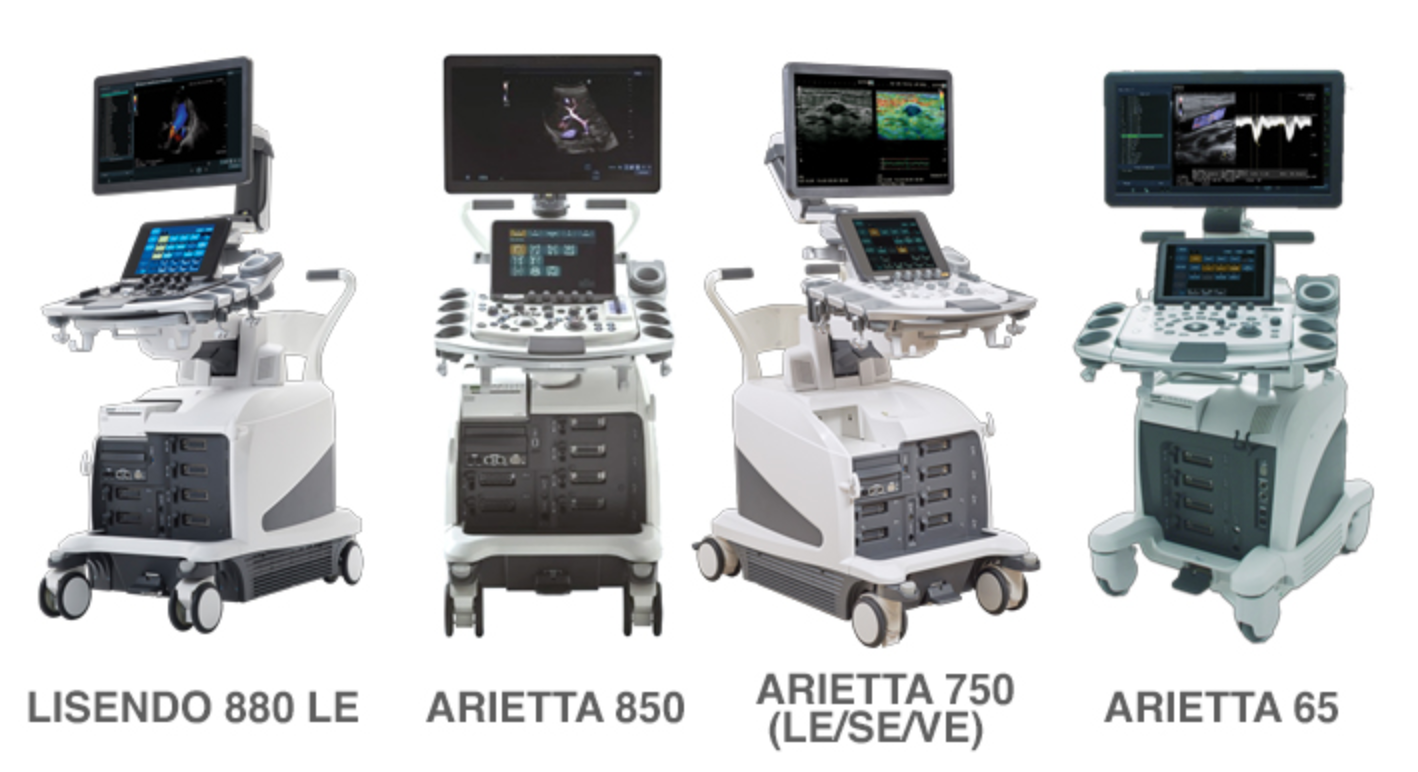
Our AI technology is integrated in all of our products.
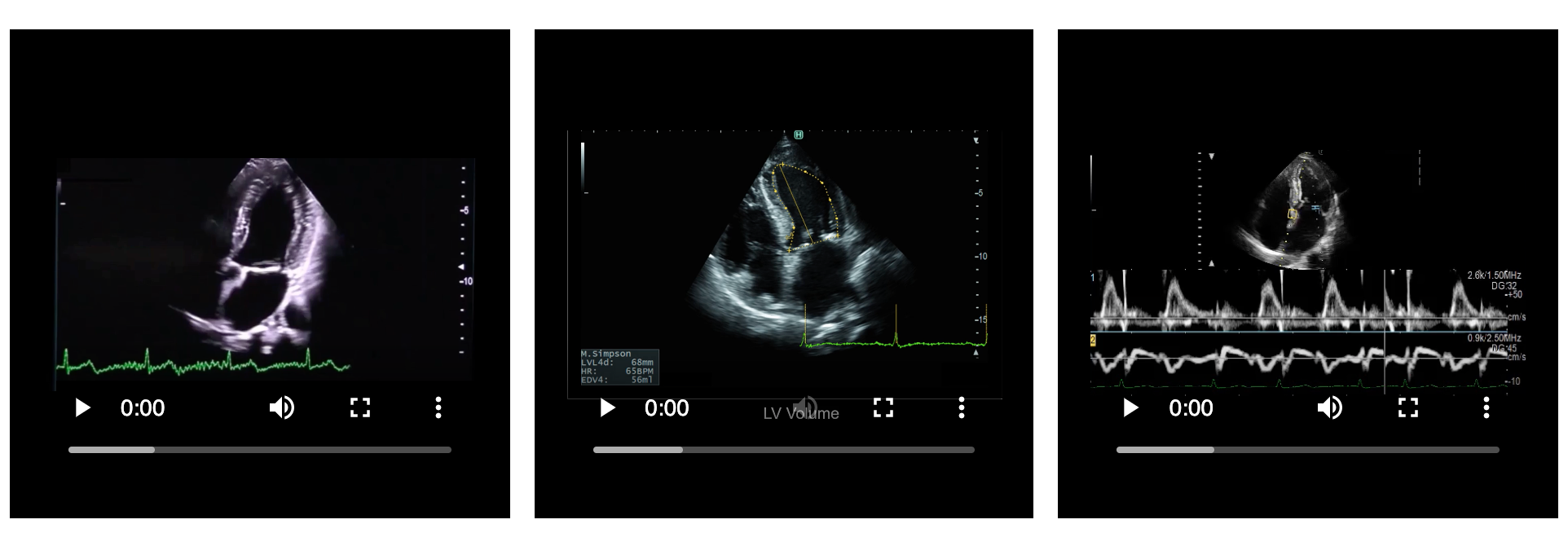
Automated Ed-Es detection
Our AI technology automatically recognizes Ed and Es phases by analyzing ECG signal after freezing the image. Ed and Es images can be assigned in a dual screen with just one-touch. This can drastically reduce your process steps when calculating the Ejection Fraction index.
Automated Tracing
This application saves many steps when tracing the endocardium and epicardium for volume, strain, and fractional area change measurements of the left ventricle (LV), left atrium (LA), right ventricle (RV), and right atrium (RA). We tested the recognition for approximately 2000 samples including end-diastole and end-systole of A4C collected from clinical sites. Automated traces were well correlated with manual traces. When using the modified Simpson’s method, the automated tracing with manual compensation reduced the measurement time to half of fully manual tracing.
Automated Stable R-R interval detection
Manually selecting stable R-R intervals can be challenging. In patients with an irregular heartbeat, for example, in atrial fibrillation or arrhythmia related to heart failure, R-R Navigation automatically detects heartbeats appropriate for the measurement, selecting a heartbeat where the R-R interval is the same as in the previous 2 waveforms.
Doppler Casual Assist
This application sets the Doppler cursor at the position of LV inflow, outflow, or annulus automatically in PW or TDI-PW mode. In the Dual Gate Doppler mode, two Doppler cursors can be set automatically at the same time. The Deep Learning algorithm extracts the features from thousands of images including A2C, A3C, and A4C views. Cursor positions and image intensities are also learned. This learned data is stored in the ultrasound machine. Next, the machine classifies it according to a standard view automatically in reference to the learned data. And then, the cursor positions are detected at the position of LV inflow and outflow for PW, and annulus for TDI-PW in the A3C view.
Intelligence Dual Gate Doppler
Dual Gate Doppler (DGD) can obtain two Doppler waveforms simultaneously in the case of arrhythmia such as with atrial fibrillation. DGD enables users to measure E/e’and IVRT consistently. The convenience of iDGD provides the combination of Doppler Cursor Assist, R-R navigation, and Doppler auto-trace for measurements. Therefore, the combination of these automation functions improves the workflow of Dual Gate Doppler measurements. The E/e’ automated measurement consists of cardiac cycle setting based on R-R navigation, detection of E(e’) wave starting and A(a’) wave ending phases, Doppler auto-trace in detected interval, and E- and e’-wave measurement based on Doppler peak detection.
Conclusion
By developing automation functions, using machine learning, for the purpose of improving quantification and measurement efficiency, we achieved a reduction in steps for quantification and a reduction in time for performing these, frequently used functions which have been historically time-consuming, and have a high inter-observer error. Improvement of basic performance of the machine learning algorithm is an ongoing project. Future development of artificial intelligence will improve the performance and the expansion of applications.
Never stop!