According to a study
published online in American Journal of Roentgenology, there is a need to
incorporate advanced image-processing techniques and additional display methods
as they could play a role in the early detection of lung cancer during annual
screenings.
The study was conducted by Dong Ming Xu, MD and colleagues from the Department of Radiology at Mount Sinai School of Medicine in New York City. The authors suggest that a review of the varied appearance of malignant modules could prove to be beneficial.
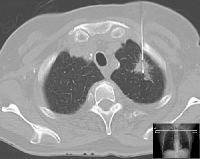
During the study, CT images of patients with lung cancer diagnosed in early rounds of screening as part of the International Early Lung Cancer Action Program (I-ELCAP) were reviewed. Scans of 104 lung cancer patients were reviewed by three radiologists. The patients were divided into three categories: patients in whom cancer was not visible at a previous CT screening; patients in whom cancer was visible but not identified at a previous CT screening; and patients in whom an abnormality was identified at a previous CT Screening but which not classified as malignant.
The results of the study showed that 77 percent of the patients could have had their cancers identified on the previous screening. These were patients who had clinical stage I disease on a prior screening image. In 18 percent of patients, by the time their subsequent annual screening occurred, their disease had already progressed beyond stage 1. The findings also showed that in 56 patients in the second category, 17 had nodules that were smaller than 3mm. Two of these patients had disease progression beyond stage I by the following year. 21 patients had nodules that were larger than 3mm and were similar in size to adjacent blood vessels. One third of these patients had their disease progress beyond stage 1 by the next annual screening.
“This finding suggests that use of computer-assisted diagnosis, which is particularly useful for separating nodules from blood vessels, would have led to even earlier diagnosis,” wrote Xu and colleagues. “Perhaps in the future such visualisation techniques will become an integral part of the reading process.”
According to the authors of this study, there is a need to use additional imaging in maximum intensity projection as it may facilitate discrimination between nodules and blood vessels. The authors also note that there is a risk of misclassifying nodules as fibrosis, scarring and simple bulla due to lack of experience in identifying small cancers with unusual appearance. They point out that distinguishing features such as convex borders, market asymmetry of apical scarring, or the presence of cystic airspace with irregularly thickened walls should alert radiologists that the patient may need follow up.
Source: Health Imaging
Image Credit: Wikimedia Commons


