Radiologists in Norway are using a CT app to facilitate image reading of the most common areas of the body for bone metastasis
The spinal cord is the vital link between the brain and body, a superhighway of life-critical information protected by the bony vertebra of the spinal column.
The spinal column is the most common site for bone metastasis, when tumors spread from internal organs to the bones. Estimates indicate that at least 30% and as many as 70%of patients with cancer will experience spread of cancer to their spine. Each year, about 10,000 Americans develop primary or metastatic spinal cord tumors.
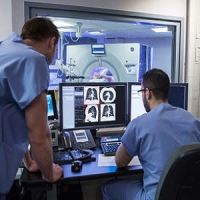
“The spine is a place where people can miss lesions, especially very small metastases, which could be the difference between finding an early, treatable tumor and finding a tumor too late to be treatable,” says Dr. Michael Fanariotis, chief radiologist for CT at the Telemark Hospital in Skien, Norway.
About 6% of CT exams is dedicated to spine imaging, but many more exams benefit from information observed within the vertebral bodies and the discs between them.
When radiologists review an imaging exam and see a lesion next to the spine, they need to precisely locate it on the image. Currently, radiologists have to manually count the vertebra to include the location of the suspicious spot in their report.
Dr. Fanariotis, is partnering with GE Healthcare to develop a new CT application that uses Deep Learning to streamline spinal imaging.
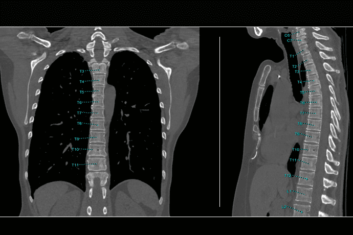
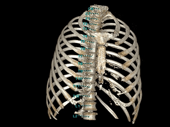
Bone VCAR is an algorithm designed to help simplify the reading experience and improve reporting efficiency by automatically labeling vertebra bodies of the spine. It can identify and label segments of the spine or the entire spine within a few seconds.
“It’s so easy. You just click a button on the toolbar and after a few seconds, you get automatic labelling of the spine simultaneously on all planes and 3D views on all your loaded CT series,” says Dr. Fanariotis. “Automatic labeling is such a big benefit because you don’t have to count again. You know exactly where you are.”
Bone VCAR helps radiologists reduce their time-consuming tasks of identifying and labeling the vertebral bodies to read scans more efficiently. It is based on Deep Learning, which uses large volumes of data sets to help the algorithm learn to recognize vertebrae in different situations. This enables spine labelling to be precise and reliable.
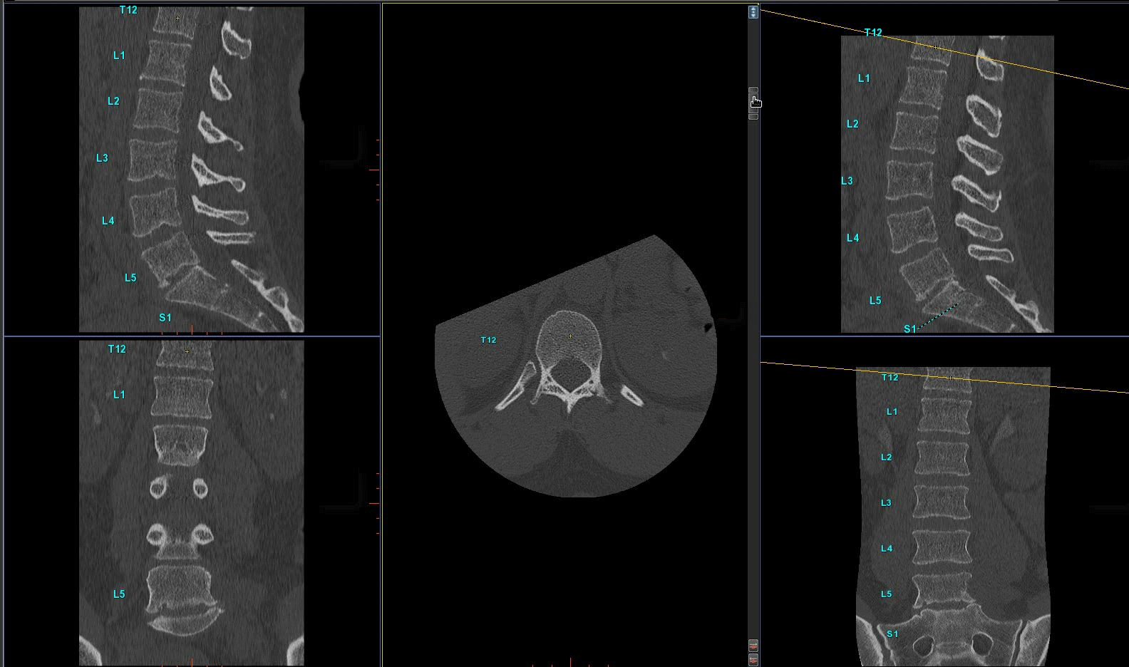
The algorithm also automatically generates the curved reformats of the spine with perpendicular oblique views to easily see the true cross section of the vertebral bodies and disc spaces, which is especially important for exams of patients with diseases like scoliosis. Previously, Dr. Fanariotis had to manually adjust the planes every few seconds to read the entire spine.
“Manually adjusting the plane every time is disrupting”, he said. “Viewing true axial planes rendered automatically as we browse through the spine helps in keeping us focused on the clinical findings. It makes reading of the spine easier.”
He says the automatic labeling and reformatting will save him approximately two to five minutes per exam – which adds up to over one hour per day.'
“It pays off at the end of the day, but it’s more than just the math,” he said. “We want to use our energy in the most effective way to reach a precise and accurate diagnosis. Because the most important resources we have are time and our concentration.”
510(k) pending at FDA. Not available for sales in the United States.