The combination of positron emission tomography (PET) and magnetic resonance imaging (MRI) has great potential. However, the clinical benefits it may bring remain unclear and need to be addressed, according to Professor Heinz-Peter Schlemmer, head of the radiology department at the German Cancer Research Centre in Heidelberg, Germany, who presented at the European Congress of Radiology in Vienna.
The interest in obtaining both morphological and functional information is great, and researchers have worked extensively on how to assess functional anatomy for about three decades, said Schlemmer ahead of the dedicated New Horizons Session on Saturday.
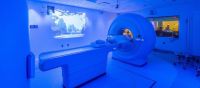
“The first milestone towards combined anatomical and functional imaging was the union of PET and computed tomography (CT) in a so-called PET-CT hybrid system in 2000. MRI provides high spatial and contrast resolution and functional information without radiation exposure. The development of a fully integrated PET-MRI system was technologically very challenging and its first use for imaging the human brain took place at the end of 2006. Whole-body PET-MRI is now commercially available and of particular medical interest for neurological, cardiovascular and oncologic diseases,” he said.
For instance, the combination of PET and MRI can improve tumour detection, by allowing radiologists to see a lesion on both morphological and metabolic images with unrivalled sensitivity. Schlemmer, who has worked extensively on comparative studies of whole-body MR and PET-CT for the detection of solid tumours, found that, while all modalities are useful in this application, depending on the particular tumour entities, MR and PET have the highest accuracy.
“Interestingly the important information came either from MR or PET studies, but CT was often not that relevant. We thought that the combination of PET and MR would be ideal to detect and typify tumours. Locally, the tumour can be imaged with the best anatomical resolution by MR, and in addition you have this metabolic and membrane receptor information about cells from PET, so together you increase your sensitivity and specificity for tumour detection and characterisation,” he said.
Nevertheless, it remains to be seen if the simultaneous acquisition of MR and PET studies will bring more accuracy than subsequent sequencing. Further information is also needed as to whether MR-PET presents added value compared to the already established PET-CT technology, and, if so, for which applications.
“Initial euphoria regarding the medical prospects of PET-MRI for assessing various diseases is understandable as this novel technology matures. But criticism is still justified, because the novel imaging technology cannot enter routine clinical practice before diagnostic accuracy, influence on therapeutic management and economic factors have been carefully considered and evidenced by clinical studies,” Schlemmer said. Capabilities and difficulties of PET-MRI have to be thoroughly balanced by considering different perspectives including scientific, medical and economic aspects, he concluded.
Latest Articles
MR-PET
The combination of positron emission tomography (PET) and magnetic resonance imaging (MRI) has great potential. However, the clinical benefits it may bring...