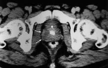
The role of magnetic resonance imaging (MRI) techniques in the diagnosis of prostate cancer has been clarified by the findings of several recently published papers. Much evidence has emerged on how methods such as MRI in combination with MR spectroscopy can improve rates of cancer detection in men with elevated levels of prostate-specific antigen (PSA) but negative biopsies, and how these techniques can help with cancer staging and treatment planning.
Currently, men with elevated PSA levels typically undergo transrectal utrasound (TRUS)-guided biopsy to determine if cancer is present. Although the results are not always definitive however it is possible that diagnostic accuracy could be increased by the use of MRI alone or in combination with MR spectroscopy. MR spectroscopy provides information on specific metabolites, such as choline (levels of which are increased if cancer is present) or citrate (levels of which are reduced if cancer is present). In a review paper published last year, researchers summarised published data on the efficacy of MRI or combined MRI/MR spectroscopy, compared with repeat biopsy, in detecting prostate cancer in men with elevated PSA and negative TRUS-guided biopsies. They evaluated six studies, with a total of 215 patients, in which the overall cancer detection rate according to the repeat biopsy result was 21-40%. With MRI or combined MRI/MR spectroscopy, the sensitivity for predicting positive biopsies was 57-100%, while the specificity was 44-96% and the accuracy was 67-85%.
In the five studies in which clinicians took MRI-targeted biopsies and standard cores, cancer was detected in 34 of 63 patients (54%) on the basis of the MRI-targeted cores only. The authors of the review concluded: “The value of endorectal MRI and MR spectroscopy in patients with elevated PSA levels and previous negative biopsies to target peripheral zone tumors appears to be significant.”
In one prospective study of 54 men with elevated PSA levels and negative TRUS-biopsies, rigorous investigation and follow-up biopsy showed that cancer was present in 17 of the men (31.5%). The combination of MRI and MR spectroscopy correctly indicated the cancer sites in all 17 of the patients, while MRI alone and MR spectroscopy alone identified cancer in 15 of these 17 patients. The researchers acknowledged that larger trials are needed, but concluded from their overall findings that MRI alone could be useful for selecting men in whom further biopsies are unnecessary, while the combination of MRI and MR spectroscopy “might be able to drive biopsies in suspicious sites and increase the cancer detection rate”.
Reviewing both the clinical and technical aspects of prostate MRI and MR spectroscopy in a paper published in June, specialists concluded that the methods are “useful tools” for the detection, localization, staging and functional assessment of the disease. As always, further work and larger studies are needed in order to improve our knowledge of how best to make use of these evolving techniques.


