


A groundbreaking imaging study published in PLOS Biology shows that the spinal cord engages in its own learning of motor tasks independent of the brain. The new findings may provide new opportunities for rehabilitation after spinal cord injury, according to researchers..
Using a 3.0T MRI scanner, the researchers performed functional magnetic resonance imaging (fMRI) of both the brain and cervical cord in subjects doing a complex finger tapping task. MRI scans revealed that learning-related changes in blood flow in the spinal cord were independent of changes in blood flow in the brain regions involved in the task.
Learning a complex motor task, such as touch typing or playing the piano, induces changes in the brain, which can be monitored using fMRI. During learning, sensory information and motor commands pass through the spinal cord. Up until now, performing fMRI on the brain and spinal cord simultaneously has been a challenge to clinicians and researchers. Hence, it has been difficult to ascertain whether observed changes in the spinal cord during motor skill acquisition depend entirely on signals from the brain, or occur independently.
That barrier was overcome for the first time in this study by taking advantage of the fact that the 3.0T MRI scanner had a field of view long enough to image the brain and the cervical spinal cord, which relays signals to and from the hand muscles. This innovative study was conducted by Shahabeddin Vahdat, Ovidui Lungu, and principal investigator Julien Doyon, of the University of Montreal, Quebec, Canada.
The results of the study indicate that the spinal cord plays an active role in the very earliest stages of motor learning. The research team says more work is needed to confirm that the changes seen in the spinal cord persist over time and generalise to other stages of learning and other forms of motor skills.
Top image: Neural correlates of motor sequence learning.
Distinct cortical, subcortical, and spinal clusters showed learning-related modulation in activity only during the CS condition. All clusters of activation are positively correlated with the performance speed. At the cortical level, the activation cluster was located in the contralateral sensorimotor cortex. At the subcortical level, one cluster was found in the contralateral putamen, while the other was observed in the ipsilateral lobule V-VI of the cerebellum. In the spinal cord, activation clusters were centred on the C7–C8 spinal segments, similar to those observed in the main effect of practice.
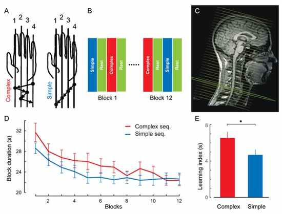
Figure 1. Behavioural and imaging protocols.
(A) The complex (CS; 4-1-3-2-4) and simple (SS; 4-3-2-1) motor sequence learning tasks were executed with the left (nondominant) hand. Subjects were required to execute 12 CS and 12 SS blocks of practice, with 60 movements each. (B) The CS and SS conditions were split evenly across blocks and alternated in a pseudorandom fashion. A 15-s rest period preceded and followed each block. (C) Functional axial slices (displayed here over the anatomical image of a representative subject) were acquired and covered both brain and cervical spinal cord up to the first thoracic (T1) segment, and they were placed at an angle that was perpendicular to the C4 vertebral segment. (D) Performance speeds (ie, block duration) averaged across all subjects show that the learning curves differed between the CS (red) and SS (blue) conditions. Participants reached asymptotic performance after the fourth block in the SS and after the eighth block in the CS condition. (E) Learning index (mean duration of the last two blocks subtracted from the first two blocks’ mean) revealed a significant difference in performance between the CS and SS conditions. Error bars represent standard error of the mean (SEM); * indicates p<0.05.
Source and image credit: PLOS
Using a 3.0T MRI scanner, the researchers performed functional magnetic resonance imaging (fMRI) of both the brain and cervical cord in subjects doing a complex finger tapping task. MRI scans revealed that learning-related changes in blood flow in the spinal cord were independent of changes in blood flow in the brain regions involved in the task.
Learning a complex motor task, such as touch typing or playing the piano, induces changes in the brain, which can be monitored using fMRI. During learning, sensory information and motor commands pass through the spinal cord. Up until now, performing fMRI on the brain and spinal cord simultaneously has been a challenge to clinicians and researchers. Hence, it has been difficult to ascertain whether observed changes in the spinal cord during motor skill acquisition depend entirely on signals from the brain, or occur independently.
That barrier was overcome for the first time in this study by taking advantage of the fact that the 3.0T MRI scanner had a field of view long enough to image the brain and the cervical spinal cord, which relays signals to and from the hand muscles. This innovative study was conducted by Shahabeddin Vahdat, Ovidui Lungu, and principal investigator Julien Doyon, of the University of Montreal, Quebec, Canada.
The results of the study indicate that the spinal cord plays an active role in the very earliest stages of motor learning. The research team says more work is needed to confirm that the changes seen in the spinal cord persist over time and generalise to other stages of learning and other forms of motor skills.
Top image: Neural correlates of motor sequence learning.
Distinct cortical, subcortical, and spinal clusters showed learning-related modulation in activity only during the CS condition. All clusters of activation are positively correlated with the performance speed. At the cortical level, the activation cluster was located in the contralateral sensorimotor cortex. At the subcortical level, one cluster was found in the contralateral putamen, while the other was observed in the ipsilateral lobule V-VI of the cerebellum. In the spinal cord, activation clusters were centred on the C7–C8 spinal segments, similar to those observed in the main effect of practice.
Figure 1. Behavioural and imaging protocols.
(A) The complex (CS; 4-1-3-2-4) and simple (SS; 4-3-2-1) motor sequence learning tasks were executed with the left (nondominant) hand. Subjects were required to execute 12 CS and 12 SS blocks of practice, with 60 movements each. (B) The CS and SS conditions were split evenly across blocks and alternated in a pseudorandom fashion. A 15-s rest period preceded and followed each block. (C) Functional axial slices (displayed here over the anatomical image of a representative subject) were acquired and covered both brain and cervical spinal cord up to the first thoracic (T1) segment, and they were placed at an angle that was perpendicular to the C4 vertebral segment. (D) Performance speeds (ie, block duration) averaged across all subjects show that the learning curves differed between the CS (red) and SS (blue) conditions. Participants reached asymptotic performance after the fourth block in the SS and after the eighth block in the CS condition. (E) Learning index (mean duration of the last two blocks subtracted from the first two blocks’ mean) revealed a significant difference in performance between the CS and SS conditions. Error bars represent standard error of the mean (SEM); * indicates p<0.05.
Source and image credit: PLOS
References:
Vahdat S, Lungu O, H, Doyon J et al.
(2015) Simultaneous Brain-Cervical Cord fMRI Reveals Intrinsic Spinal Cord Plasticity during Motor Sequence Learning. PLoS Biol, 2015 DOI:
10.1371/journal.pbio.1002186
Latest Articles
healthmanagement, brain, spinal cord, motor skills, fMRI, learning
A groundbreaking imaging study published in PLOS Biology shows that the spinal cord engages in its own learning of motor tasks independent of the brain.

 innervates 5 dendritic spines (orange, labeled 1–5)")

")


