


A new study published in Radiology reveals that degeneration of the brain's white matter may be an early marker of specific types of Alzheimer’s disease (AD), including early-onset AD. Researchers used diffusion tensor imaging (DTI) to assess the white matter tracts in 53 patients with three types of AD: early-onset AD and two atypical types of AD called focal syndromes because they affect localised parts of the brain.
“Alzheimer’s is a grey matter disease,” notes Federica Agosta, MD, PhD, co-author of the study conducted at the Neuroimaging Research Unit, San Raffaele Scientific Institute in Milan, Italy. “However, white matter damage has a central role in how the disease strikes and progresses.”
AD is an irreversible, progressive brain disease that slowly destroys memory and thinking skills. Where or how the disease begins is not fully understood. Unlike late-onset AD that occurs after age 65 and is characterised primarily by progressive memory loss, patients with early-onset AD have impairment in several regions of the brain, including deficits in executive functioning and visuospatial abilities. Focal AD syndromes may cause visual disturbances or language deficits.
Researchers used DTI to identify similarities and differences in white tract damage across the AD spectrum and in relation to patterns of cortical atrophy, according to Dr. Agosta. DTI is a specialised magnetic resonance imaging technique that uses the movement of water molecules to characterise the microstructure of biological tissues. The technique is highly sensitive to white matter degeneration, she says.
Analysis of the images revealed that all of the patients had extensive white matter damage, and showed regional grey matter damage. “The white matter damage in patients with focal AD syndromes was much more severe and widespread than expected and cannot be explained solely by grey matter atrophy which was more localised,” Dr. Acosta points out.
She adds that the findings support the theory that AD pathology may travel along white matter fibres from one region of the brain to another.
In early-onset AD and atypical AD forms, "white matter degeneration may be an early marker that precedes grey matter atrophy,” Dr. Agosta says, adding that DTI has the potential to assess the extensive disorganisation of brain networks in focal AD even before overt cognitive deficits become apparent.
Top image.MR images of A, common and B, syndrome-specific patterns of cortical atrophy across the Alzheimer's disease variants.
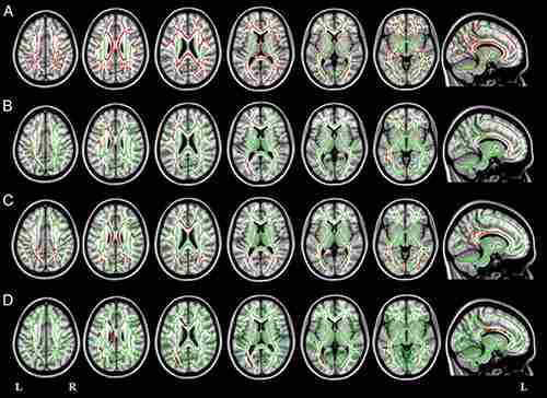
Figure 2. Axial (left six columns) and sagittal (right column) MR images of fractional anisotropy results in patients with Alzheimer's disease variants relative to matched healthy control subjects.
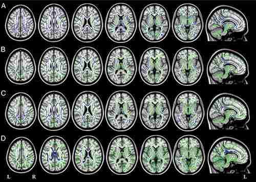
Figure 3. Axial (left six columns) and sagittal (right column) MR images of mean diffusivity results in patients with Alzheimer's disease variants relative to matched healthy control subjects.
Source and image credit: RSNA
“Alzheimer’s is a grey matter disease,” notes Federica Agosta, MD, PhD, co-author of the study conducted at the Neuroimaging Research Unit, San Raffaele Scientific Institute in Milan, Italy. “However, white matter damage has a central role in how the disease strikes and progresses.”
AD is an irreversible, progressive brain disease that slowly destroys memory and thinking skills. Where or how the disease begins is not fully understood. Unlike late-onset AD that occurs after age 65 and is characterised primarily by progressive memory loss, patients with early-onset AD have impairment in several regions of the brain, including deficits in executive functioning and visuospatial abilities. Focal AD syndromes may cause visual disturbances or language deficits.
Researchers used DTI to identify similarities and differences in white tract damage across the AD spectrum and in relation to patterns of cortical atrophy, according to Dr. Agosta. DTI is a specialised magnetic resonance imaging technique that uses the movement of water molecules to characterise the microstructure of biological tissues. The technique is highly sensitive to white matter degeneration, she says.
Analysis of the images revealed that all of the patients had extensive white matter damage, and showed regional grey matter damage. “The white matter damage in patients with focal AD syndromes was much more severe and widespread than expected and cannot be explained solely by grey matter atrophy which was more localised,” Dr. Acosta points out.
She adds that the findings support the theory that AD pathology may travel along white matter fibres from one region of the brain to another.
In early-onset AD and atypical AD forms, "white matter degeneration may be an early marker that precedes grey matter atrophy,” Dr. Agosta says, adding that DTI has the potential to assess the extensive disorganisation of brain networks in focal AD even before overt cognitive deficits become apparent.
Top image.MR images of A, common and B, syndrome-specific patterns of cortical atrophy across the Alzheimer's disease variants.
Figure 2. Axial (left six columns) and sagittal (right column) MR images of fractional anisotropy results in patients with Alzheimer's disease variants relative to matched healthy control subjects.
Figure 3. Axial (left six columns) and sagittal (right column) MR images of mean diffusivity results in patients with Alzheimer's disease variants relative to matched healthy control subjects.
Source and image credit: RSNA
Latest Articles
healthmanagement, brain atrophy, Alzheimer's disease, biomarkers, white matter damage
A new study published in Radiology reveals that degeneration of the brain's white matter may be an early marker of specific types of Alzheimer’s disease (AD), including early-onset AD.


