Peritoneal metastases remain one of the harder problems in abdominal oncology imaging. Small deposits can be difficult to distinguish from surrounding soft tissue, yet missing them can alter staging, surgical selection and the extent of treatment. A new study published on 30 September in European Radiology Experimental examines whether spectral photon-counting CT can improve that visibility when reconstruction is tuned specifically for the task.
Why small peritoneal lesions remain difficult
CT is central to the preoperative assessment of colorectal cancer, but its sensitivity falls sharply as peritoneal deposits become smaller. The authors cite previous evidence reporting sensitivity of about 11% for nodules below 5 mm and 25–50% for lesions under 1 cm. Low soft-tissue contrast is part of the problem. Spectral imaging can increase contrast at low virtual monoenergetic energies, but those images may also become noisier and more heavily smoothed.
That trade-off is precisely what the new study tested. Rather than asking whether photon-counting CT is generally “better”, the investigators looked at which reconstruction settings best balance noise, spatial resolution and lesion detectability for peritoneal disease.
Matched-dose comparison of photon-counting and dual-energy CT
The team used a spectral phantom containing iodine-based inserts selected to reproduce the contrast expected from mucinous and non-mucinous peritoneal metastases on 40 keV virtual monoenergetic images. The phantom was scanned on both a spectral photon-counting CT research system and an energy-integrating dual-energy CT system at closely matched radiation exposure, with a CTDIvol of approximately 8.5 mGy.
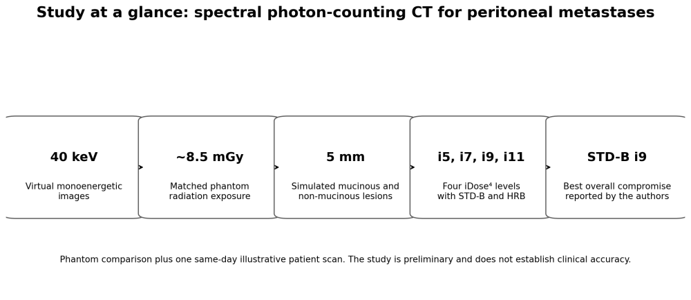
Editorial visualisation based on Greffier et al., European Radiology Experimental 10, 144 (2026). Source data: 40 keV VMIs, matched phantom CTDIvol of about 8.5 mGy, 5 mm simulated detection tasks, and four iDose⁴ levels.
For photon-counting CT, the researchers tested two reconstruction kernels and four iterative reconstruction levels. They then measured noise power, spatial resolution and a task-based detectability index for simulated 5 mm lesions. This matters because a reconstruction that looks smoother is not automatically the reconstruction that preserves the small structures radiologists need to see.
STD-B with iDose⁴ level 9 gave the best overall balance
The main result was a clear reconstruction trade-off. Increasing iterative reconstruction reduced noise, but at the highest settings it also began to soften lesion margins. Across the combinations tested, the STD-B kernel at iDose⁴ level 9 provided the best overall compromise between noise texture, spatial resolution and detectability.
With the standard kernel, detectability for both simulated mucinous and non-mucinous metastases was similar to or better than conventional dual-energy CT from iterative levels 7 through 11. The photon-counting system also produced finer noise texture and, for most of the optimised settings, slightly better spatial resolution. For clinical adoption, performance therefore depends on the reconstruction pipeline as well as the detector. Reconstruction choices can materially change what the examination reveals.
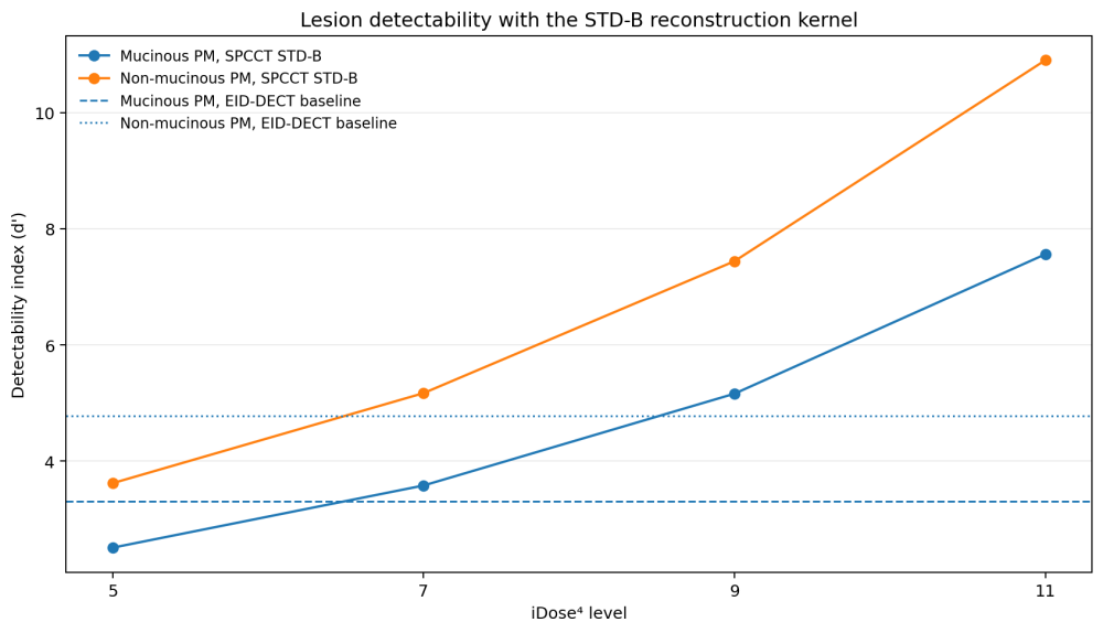
Editorial chart based on Table 2. With STD-B, d' was below the EID-DECT baseline at i5, then higher from i7 onwards for both simulated lesion types. EID-DECT baselines are derived from the published relative differences and rounded to two decimals.
A patient example shows the clinical direction
The investigators also included an illustrative same-day comparison in a 59-year-old woman with synchronous peritoneal metastases. Radiation exposure was closely matched between the two systems, at 8.2 mGy for dual-energy CT and 8.3 mGy for photon-counting CT. On the photon-counting images, the authors reported improved delineation of muscular fascia and clearer visualisation of a peritoneal nodule and omental fat stranding with the better-balanced reconstruction settings.
This patient example is useful, but it is not a clinical accuracy study. The formal comparison was phantom-based, with one illustrative patient and no statistical analysis. The paper therefore does not establish that photon-counting CT improves staging accuracy, changes treatment decisions or improves outcomes in patients with colorectal cancer.
Why this matters for oncology CT
For imaging leaders, the relevance is broader than one abdominal application. Photon-counting CT is increasingly discussed in terms of detector efficiency, spectral information and ultra-high resolution. This study shows the next implementation challenge: converting those technical capabilities into task-specific protocols that preserve clinically important detail rather than simply generating attractive images.
Peritoneal disease is a useful test case because the lesions are often small, low contrast and consequential. If future clinical studies confirm that optimised photon-counting protocols improve detection and staging, the impact could extend directly into surgical planning and patient selection. For now, the evidence should be read as technically encouraging rather than clinically definitive.
Study context: Both CT systems were Philips Healthcare platforms. One co-author, Maria Nicole Antonuccio, is an employee of Philips Healthcare; the authors reported no other relevant conflicts of interest or industry support for the project.
Source: Springer Nature
Image Credit: iStock
References:
Greffier J, Grange R, Dabli D, et al. Optimization of spectral photon-counting CT reconstruction parameters for improved detection of peritoneal metastases compared with energy-integrating detector dual-energy CT: a phantom study. European Radiology Experimental. 2026;10:144. Published 30 September 2026.