Photon-counting CT first gained attention in cardiovascular imaging, where spatial resolution, spectral information and reduced blooming offered an obvious advantage in calcified coronaries and stented vessels. A Siemens Healthineers collection of 13 clinical cases now illustrates a much broader pattern of use. The cases cover abdominal, cardiovascular, musculoskeletal, neuro and head-and-neck imaging, with clinical questions ranging from pancreatic cancer and an occult fracture to cerebrospinal fluid leakage and intracranial aneurysm follow-up.
One detector, very different clinical questions
The value of the collection is not that 13 cases prove superiority. They do not. Case reports are descriptive and results from one institution cannot be assumed to transfer to another. What the collection does show is the range of problems for which clinicians are applying photon-counting CT in routine practice.
In Budapest, a patient presenting with haemoptysis, dyspnoea and chest pain underwent a single ECG-gated chest angiogram on NAEOTOM Alpha.Prime. The examination demonstrated pulmonary embolism with infarct pneumonia, assessed the coronary arteries despite an irregular heart rate and used spectral lung analysis to map the perfusion deficit. The case revisits the triple-rule-out concept, but with the spectral and temporal capabilities of current photon-counting CT.
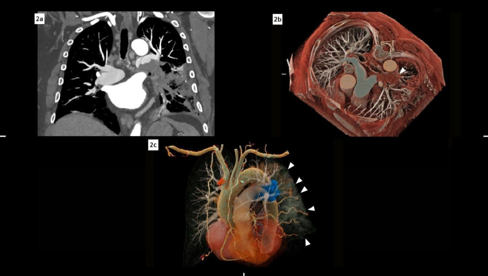
Figure 1. Triple-rule-out PCCT. Coronal MIP and VRT reconstructions demonstrate a left pulmonary artery filling defect; the oblique VRT also shows left-lung consolidations.
Credit: Semmelweis University, Medical Imaging Center, Budapest, Hungary / Siemens Healthineers.
Oncology: resolving uncertainty that affects the pathway
A pancreatic ductal adenocarcinoma case from Peking Union Medical College illustrates a different use. Conventional CT had shown a pancreatic body tumour but left two important uncertainties: possible intraductal tumour spread and a small hepatic lesion that could represent metastasis.
Photon-counting CT with ultra-high-resolution acquisition and 50 keV virtual monoenergetic images more clearly defined the tumour. The suspected intraductal extension was not supported, while the small liver lesion was characterised as a sharply delineated non-enhancing cyst. Thickening and enhancement of the left coeliac ganglion raised concern for perineural spread, which was later confirmed pathologically after surgery.
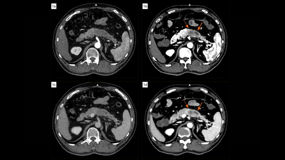
Figure 2. Pancreatic ductal adenocarcinoma. EID CT images (left) are compared with PCD CT images (right); the PCD images more clearly delineate the tumour and adjacent pancreatic duct, with VMI displayed at 50 keV.
Credit: Department of Radiology, Peking Union Medical College, Beijing, P. R. China / Siemens Healthineers.
The case is important because it moves the discussion beyond image quality. Additional spatial and spectral information matters when it resolves an ambiguity that could alter staging or surgical planning.
Musculoskeletal and neuro cases show where resolution counts
At Semmelweis University, a dens fracture that had not been visible on previous energy-integrating detector CT images acquired at 0.625 mm was clearly depicted on photon-counting CT images acquired at 0.4 mm. The authors attributed the difference to improved spatial resolution and reduced partial-volume effects.
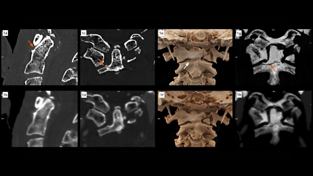
Figure 3. Dens fracture. The C2 dens fracture is depicted on PCD CT acquired at 0.4 mm after not being visible on the preceding EID CT acquired at 0.625 mm; cVRT views show the fracture anatomy.
Credit: Semmelweis University, Medical Imaging Center, Budapest, Hungary / Siemens Healthineers.
The same collection includes neurovascular follow-up after aneurysm treatment, a cerebrospinal fluid-venous fistula in a patient with a long history of headaches, traumatic middle-ear injury and cerebral lymphoma. These are very different referrals, but they share a requirement for reliable depiction of small structures, contrast differences or anatomy affected by metal and other artefacts.
The portfolio question is changing
For hospital and imaging leaders, the strategic issue is no longer whether photon-counting CT belongs in a single premium cardiac service. The more relevant question is how much of the routine and complex CT workload can migrate to a platform that combines high spatial resolution with spectral information in every scan.
That does not mean every conventional scanner should be replaced immediately. The evidence supporting PCCT varies considerably by indication, and much of the Siemens material is case-based. Procurement decisions still need workload data, service-line demand, lifecycle cost, staffing and downstream clinical evidence.
What has changed is the breadth of the clinical conversation. Photon-counting CT is increasingly being used across cardiac, oncology, emergency, musculoskeletal and neurological pathways. If that breadth is confirmed by larger comparative studies, the technology may be judged less as a specialist scanner and more as a general CT platform for high-complexity care.
Source: Siemens Healthineers
Image Credit: iStock