Study Stresses the Importance of Brain Monitoring During the Treatment of Patients with Severe COVID-19
Masimo (NASDAQ: MASI) today announced the results of a prospective, observational study published in Critical Care in which researchers in Genoa, Italy, evaluated the impact of a variety of rescue therapies on the systemic and cerebral oxygenation of mechanically ventilated COVID-19 patients suffering from acute respiratory distress syndrome (ARDS).1 To gauge the impact, the researchers used the Masimo Root® Patient Monitoring and Connectivity Platform with O3® Regional Oximetry, which uses near-infrared spectroscopy (NIRS) to enable monitoring of tissue oxygen saturation (rSO2) in the region of interest, such as the brain.
This press release features multimedia. View the full release here: https://www.businesswire.com/news/home/20210405005487/en/
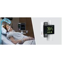
Masimo Root® with O3® Regional Oximetry and SedLine® Brain Function Monitoring (Photo: Business Wire)
Dr. Chiara Robba and colleagues noted that “neurological complications are common in mechanically ventilated critically ill patients with COVID-19 and may lead to impaired cerebral hemodynamics,” and further, that respiratory rescue therapies “may have detrimental effects on brain physiology.” Observing, however, that there is currently little data available regarding the effect of rescue therapies on these patients’ brains, and in particular on cerebral oxygenation, the researchers sought to assess the impact of different ventilatory rescue therapies on the brain to help guide clinicians in choosing the most appropriate therapies for their COVID-19 patients.
The rescue therapies studied were recruitment maneuvers (RMs), prone positioning (PP), inhaled nitric oxide (iNO), and extracorporeal carbon dioxide removal (ECCO2R). To assess impact, the researchers measured (before and after the application of each method) arterial oxygen saturation (SpO2), partial pressure of oxygen (PaO2), partial pressure of carbon dioxide (PaCO2), and cerebral oxygen saturation (rSO2). rSO2 was obtained using Masimo Root with O3, which also allowed them to observe several additional parameters unique to Masimo O3: ΔO2Hb, which monitors relative changes in the oxygenated hemoglobin component of rSO2; ΔHHb, which monitors relative changes in the deoxygenated hemoglobin component of rSO2; and ΔcHb, which monitors relative changes in total cerebral hemoglobin or blood volume. As a secondary aim, the researchers sought to evaluate the correlation between systemic and cerebral oxygenation.
The researchers found that the four rescue therapies had varied impact on cerebral oxygenation and the other measured parameters, noting in particular that after RMs, while there was no significant change in PaO2 or PaCO2, there was a significant decrease in rSO2. After PP and after iNO therapies, both PaO2 and rSO2 increased; ΔcHb also increased, corresponding to increased cerebral blood volume. After ECCO2R, both PaO2 and rSO2 decreased.
The researchers concluded, “Rescue therapies exert specific pathophysiological mechanisms, resulting in different effects on systemic and cerebral oxygenation in critically ill COVID-19 patients with ARDS. … The choice of rescue strategy to be adopted should take into account both lung and brain needs.”
They also noted, “To our knowledge, this is the first study investigating the early effects of rescue therapies on systemic and cerebral oxygenation and their correlation in critically ill patients with COVID-19-associated ARDS. The use of multimodal neuromonitoring, including new indices such as ΔHHbi + ΔO2Hbi, enabled us to better investigate the specific consequences of each ventilatory rescue strategy for brain and lung function. This is particularly important, especially in the early phases after rescue therapies application, when most of the effects on cerebral physiology are mainly acting.”
Dr. Robba and study co-author Dr. Basil Matta, Senior Medical Director at Masimo, commented, “The ability to observe relative changes in oxygenated, deoxygenated, and total hemoglobin with O3’s delta indices provided us with better insight into why brain saturations change as a result of interventions, and allowed us to better understand the interactions between systemic and cerebral hemodynamics. For example, we saw that turning patients prone resulted in improved systemic and cerebral oxygenation, whereas the lung recruitment maneuver did not improve systemic oxygenation, and even had an adverse effect by reducing brain oxygen saturation.”
They continued, “Above all, the main objective of improving the oxygen content of the blood is to deliver oxygen to vital organs, the most important of which is the brain. Masimo O3 provides the clinician with the ability to assess the impact of any medical intervention aimed at improving oxygenation. O3’s hemoglobin indices were critical to our understanding of the effects of our interventions on the brain. Without such a monitor, we are at best guessing, and in danger of flying blind. As we continue to seek to improve care and outcomes for patients with severe COVID-19, any tool that helps us better understand the impact of different medical interventions is most welcome.”


