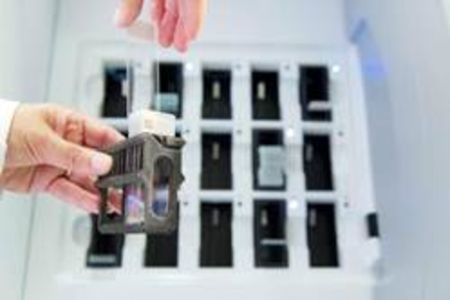
Erasmus MC is the first hospital in the Netherlands to switch to digital for their experimental laboratory analysis of cell and tissue samples using a digital pathology system from Philips. This switch to digital is an important step in tumour research and ultimately aims to speed up and improve the diagnosis and treatment of cancer and other diseases. Erasmus MC is working with Philips, which has developed a completely new, very fast technology for scanning, image processing and analysis that makes it possible to obtain digital images of suspect tissue at very high resolution. This enables medical researchers to view the images efficiently from any given workplace and to gain new insight into diseases such as cancer.
If cancer is suspected in a patient, tissue is removed surgically or by means of a biopsy. The tissue is then examined by a pathologist at microscopic level and sometimes also tested at molecular level. This makes it possible to ascertain whether or not the tissue is cancerous and, if so, to what extent the cancer is malignant. This process also plays a very important role in the analysis of large numbers of test samples for experimental cancer research in order to gain a better understanding of the causes and mechanisms of diseases at cellular and molecular level. These new insights may enable new diagnostic approaches and therapeutic treatments.
The analysis of small tissue samples can create quite a problem in medical investigations. A great deal of time and effort is spent sending, recording and processing the hundreds of microscope slides of tissue samples. Consultation with a colleague at another location can be a lengthy process, as the tissue slide first has to be sent over by courier, with the added risk of damage or loss.
Digital
By scanning the tissue slide using the very fast Philips digital pathology system, the examining pathologist can gain direct access to the digital files and the work can be distributed more effectively among the available researchers. Cancer cells in the tissue can be identified and analysed quickly using advanced image analysis software. It also becomes easy to share information and images with cancer research institutes all over the world.
"In recent years the demand for cell and tissue examination has risen enormously with more complex cases," says Peter Riegman PhD, Head of the Erasmus MC Tissue Bank. "The changeover in the future to digital pathology will help our team of pathologists, biomedical researchers, technical specialists as well as the management team to ensure we maintain our high standard and further speed up experimental medical research."
"For over a hundred years now pathologists have used an optical microscope to examine the stained tissue on a microscope slide," says Perry van Rijsingen, General Manager of Philips Digital Pathology. "By integrating digital pathology in the existing information system at the research laboratory, Erasmus MC now has at its disposal a digital platform that offers new opportunities for intensive cooperation in education and research with other disciplines such as radiology."
The Philips system for digital pathology consists of an ultra-fast scanner and image management system with software for viewing, analysing and interpreting the images. At Erasmus MC the changeover from analog to digital will be made first in experimental research and pathology education, and this may well be followed by digital diagnostics in the coming years.


